


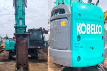


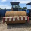
USED HEAVY MACHINERY & LORRY
RM160,000

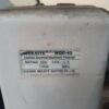
FOOD CONTAINER
RM20,000
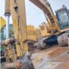
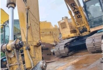
Caterpillar 320B Excavator (Magnet)
RM165,000